 |
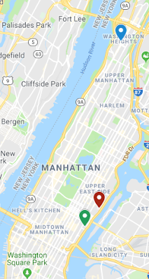
Map of locations visited this week.
Blue: Columbia Medical Center
Red: Cornell Medical Center
Green: Dr. Prince’s office.
|
One of the most enjoyable things I got to see this week was how organ volume is measured with MR images. The images would first be loaded into a workstation with a proprietary software made by the same company who makes the scanners (for some reason this could not be done on the “main” PACS dedicated computers for reading the images and required separate software on a separate workstation). Because images are acquired by sectioning the body into slices, we can determine the area of the organ on each slice and do an interpolation between the areas on all the slices to determine an approximate organ volume. This is exactly what the software would do. To make things easier, the radiologist would simply trace the outline of the organ on every 2 to 3 slices and then tell the computer to only keep the organ sections of the slices (this process is known as segmentation. Then a tool could be used in the software to measure (interpolated) the volume of what is left behind.
I really liked seeing this segmentation because it seemed simple to do. After showing me how this was done Dr. Prince let me measure the kidney, liver and spleen volumes for a few patients. Of course, he verified the measurements I made by performing them himself and used his results in any report that was generated. I was pleased that I was within +/-10% of his numbers. I did learn several things while doing this though. The first is that when cysts (or other pathologies) are present, especially in multiple organs, it can sometimes be hard to determine the border of the organ of interest. This was particularly important for me to see because Dr. Prince mentioned to me how this is one of those things that is tedious to do and should really be automated. I could see how this would be challenging for a computer to do if it was challenging for the human eye. I could however see a hybrid sort of automation as a possibility. By this I mean the computer program would draw a border on each slice where the computer “thinks” the boarder is, but the radiologist would first look at the computer chosen boarder and correct any mistakes before the volume was computed. In principle this could save 80-90% of the work.
It amazed me to learn just how many things there are in the radiologist’s day that actually could be automated to some degree that aren’t. As another example, when the radiologist reads images, they bring up two programs: one to view the images and one to create, view and assign reports. What was interesting to see is the two programs don’t talk to each other. By this I mean if you bring up the report template for a patient on the report program the images are not brought up automatically on the other program. As an engineer something can be learned here, which is that when developing software (or anything) for that matter it is important to talk to the people who will be using it, so their life is easier. I think in some sense this is what is truly great about this summer immersion. That is the ability to see firsthand how any medical technology you might make will be used.
When Dr. Prince and I were at the Columbia Medical Center I got to learn more about the “interworking” of radiologists within a hospital. With Dr. Prince I attended a liver conference on Wednesday and later that day a resident conference. Dr. Prince called the resident conference “interesting case day” as will be explained shortly. In the liver conference, a large group of radiologists would sit in a conference room and bring up patient cases on a projector that were hard to access for whatever reason (either the image was hard to read, or how the patient should be treated was tricky). Since the conference was the “liver conference” all cases involved the liver. The radiologists would all weigh in on the case and determine a “best course of action” as a group. I think this was important and good to see, because when something is hard to access it is good to get multiple opinions (especially when dealing with human life). What was the most interesting for me to learn was that many of the discussions involved patient reluctance to treatment. For example, clinically, the most sensible thing to do may be surgery, but the patient may not want to do this, and so the doctors would have to debate how and if this could be worked around.
The resident conferences were a bit different. Here the attending radiologist would set one of the residents at a PACS dedicated computer and bring up a case for the resident to look at. The resident probably had not seen the case before and so was blind to what was going on. They were then asked to read the case and the attendings would make comments. Not surprisingly many of the cases were “interesting” as Dr. Prince would put it to make the simulated situation harder. Many of the cases used came from the liver conference earlier in the day. I think that this is a great way to really learn the “art” of reading images and in some way am wondering how a simulated situation may be useful in engineering education.
One of the interesting cases that Dr. Prince brought was from a reading he made earlier in the week. This one was tricky due to an artifact. The patient had an MRA scan of the chest and one of the images that was generated was a subtraction image (literally where one image is digitally subtracted from another to help better view the anatomy of interest). On the subtraction image it looked like the patient had an aortic dissection extending from the diaphragm all the way to the iliacs. However, this pathology was not present on any of the other sequences. This made it very likely that the “pathology” we saw on the subtraction image was an artifact and not actually present. From this I learned that it is important to look at something as many ways as possible before making an assessment. Like with anything else, the more evidence you have for something existing the more likely it is to exist.
One more thing that I learned this week dealt with clinical research. I watched as Dr. Prince was viewing a bunch of research cases looking for stenosis of the IVC due to renal or hepatic cysts (as written about in my week 2 blog). Dr. Prince was going back over cases that he had already read to make sure they were read correctly as the data he was collecting was mostly based on judgement calls made by the reading radiologist. He emphasized how important this was to make sure the data (and the statistics) are accurate. I would have never thought to do this, but It makes sense when the data is not simply measurements. During this process he also saved many images to use in his report. These images showed what the statistics simply would not and painted a picture of some interesting things going on. For example, a few of the images showed how the right kidney could be pinching the IVC, but because of a cystic liver that pushed the kidney into the IVC.

No comments:
Post a Comment