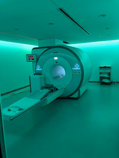
The outpatient imaging center has two MR devices, both with 3T scanner manufactured by Siemens. Before my visit to the scanner, I completed the online MR safety training class by New York-Presbyterian Hospital. The MR scanning practice is conducted in the specific place which can be classified into four areas. Zone 1 and 2 are usually for patients to wait and be notified with any clinical or safety-related concerns. Zone 3 is the control room where technicians operate the machine and Zone 4 is the MR scanning room located the machine and other appliances for examination. Patients come to the front desk for inquiry of their exam orders by appointment. Then they wait in the waiting room outside of the scanning room which is decorated with care until they are called by physicians to get into the examination areas. Before the scanning, patients are being asked with any safety-related concerns in different ways for several times, in terms of any ferromagnetic materials, implants with MR compatibility, etc. After that, patients are required to change their clothes, enter into the control room, again communicate with the technicians before they finally get into the scanning room.
 |
| 3T PET/MR scanning room at Weill Cornell Imaging |
 |
| 3T MR scanning room at Weill Cornell Imaging |
During the scanning, technicians will exam the result in order to do any post-processing protocols using the platform provided by Siemens. The platform provides a great variety of functions for post-processing. In terms of the PET/MR, the fusion images of both attenuation correction maps by PET and different weighted images by MR are made prior before sending the results to radiologists for reading and examination. However, during my visit, an unexpected artifact arose when the AC map was calculated for PET/MR fusion images, where a huge circular artifact occurred in several slices in the air area where there should be no signals. After several efforts using different source images together with different tissue type filters, this artifact was finally removed from the offline attenuation-coefficient map and fusion images were corrected. Other post-processing also included brain vessel 3D segmentation for MR angiography during my visit.
Another interesting observation was for the contrast MR exam. Gad-MR is commonly used for contrast enhancement for T1 weighted imaging for vasculature and other parts. The whole injection process is automatically monitored during the scan. Usually pre-contrast and post-contrast imaging are both required for comparison. Gad contrast takes effect for not a long time. The automated syringe guarantees precise timing and controlled rate of injection.
It was really excited for me to see the whole scanning processes. Communicating with the technicians as well as observing how they work greatly inspired me to think about how my own research related with MR imaging clinics. Most of the work involves post-processing which is tightly related with my work, with the only difference that I use general MATLAB or Python to do image analysis and signal processing while technicians use the commercial software by the imaging company such as Siemens. However, it tells me to take care of any post-processing procedure, when making it commercialized or applied to hospital, thinking about any possible condition and giving corresponding solution to verify the algorithm robustness as well as the compatibility.

No comments:
Post a Comment